Cells are organized into one of the 2 types of tissues:
1) epithelial tissue: cells directly connected to each other — form our skin and coat our organs
2) connective tissue: cells dispersed with extracellular matrix — make up muscle cells, nerve and immune cells
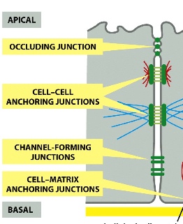
from top to bottom:
- occluding junctions: seal the space in between cells with tight junctions
- cell-cell anchoring junctions: actin or intermediate filaments of two cells are jointed together using various proteins
- channel forming junctions: proteins form channels and allow molecules to go through
- cell-matrix anchoring junctions: basal lamina is connected to the basal side of the cell with anchors
Adherens junction structure:
- actin filaments are involved
- can form strong adherens belts between epithelial cell to hold them together
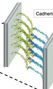
- adhesion is achieved by cadherin clusters from both sides of the two cells - homophilic interactions (meaning that one type of cadherin only interact with cadherins of the same type on the other side)
- actin filaments link to adaptor proteins link to extracellular cadherin proteins

- without adherens, tissues can disintegrates due to the lack of adhesion
- adherens junctions maintain the proper organizations of cells (which are lost during cancer development), therefore cadherins are tumour suppressors — the loss of epithelial structure is a hallmark of cancer
Epithelial structure
the apical surface faces the organ lumen (i.e. inside of gut)
the basal surface faces the underlying tissue (i.e. basal lamina) —> since the two sides are different, the tissue is polar
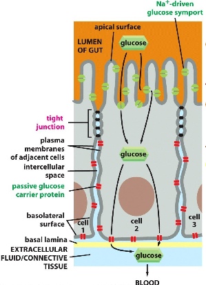
Example 1: transport of glucose
Glucose cannot pass through the space between cells. They are forced to pass through plasma membrane channels (importers and exporters on the apical and basal sides). Tight junctions not only seal the space in between (gates function), but also prevent channels from diffusing to the wrong part of the membrane (fence function; i.e. all importers are on the apical side, exporters on the basal side).
Example 2: testing the permeability barrier
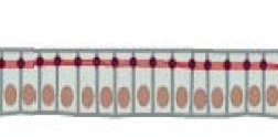
Add a dye to a monolayer of cells and see where the dye diffuses. Add tracer molecules from the apical side and also from the bask side —> all stop at the same point. Result: dye only diffuses up until the red line (actin belts) which indicates where the barrier is.
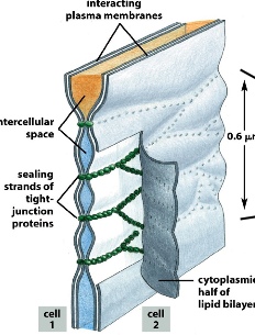
tight junctions involve many strands of transmembrane proteins interaction with each other. More specifically, each tight junction is made up of various claudins and occludins. They each has 4 transmembrane domains. Claudins are important for tight junction formation, whereas occluding are crucial for barrier composition (plugging holes).
We already know that cell polarity is important, so how is cell polarity established?
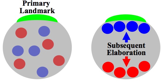
A: cells use landmarks, followed by elaboration
Landmark makes one side different from the other side —> cell recognizes it and organize intracellular materials based on this landmark.
E.g. 1: chemoattractants only bind to one side of the cell —> bind receptors —> begins actin rearrangement so that the side with chemoattractants grow (to chase after bacteria) and the side opposite of chemoattractants shrinks.
E.g. 2 in C. elegant, the side where the sperm enters the egg becomes the posterior side, and induces cytoskeleton flow so that actin and myosin go to the anterior side. This is how the embryo becomes polarized and the different regions can form different tissues later on.
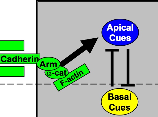
E.g. 3. Adherins can also become landmarks for epithelial polarity. Adherins are located on the apical side, and it recruits apical cues. Apical cues inhibits basal cues and vice versa.
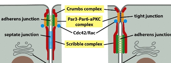
Crumbs complex and Par3-Par6-aPKC complexes are apical that define the apical domain. Scribble complex is basal.
